
Figure 4. Characterization of slow dividing NG2+ glioma cells in early hyperplasic lesions in the rostal migratory stream.
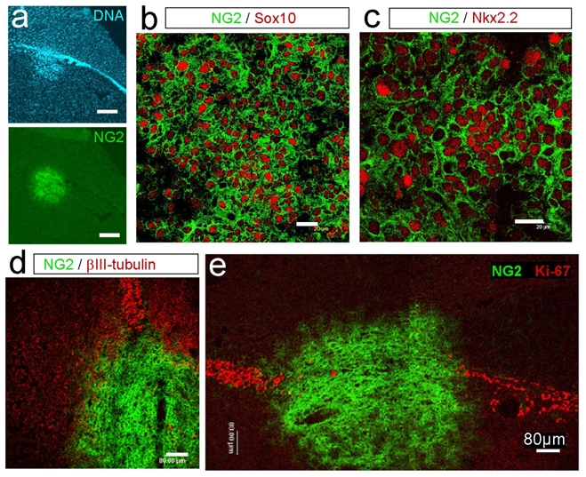
a: Brain sections of 3-month old rats stained with NG2 antibodies and counterstained with Hoechst (DNA) to visualize emerging ENU-induced tumors in the RMS, Bar=200μm.
b–c: Confocal analysis of low-grade glioma located within the RMS and double stained for NG2 (green) and Sox 10 (red) (b; Bar=20μm), NG2 (green) and Nkx2.2 (red) (c; Bar=20μm),
d–e: Confocal analysis of a low-grade glioma located within the RMS and double stained for NG2 (green) and βIII-tubulin (red) or for NG2 (green) and Ki-67 (red) (d–e; Bar=80μm).