
Figure 5. Immunocytochemical characteristics of rat low-grade glioma-derived explants.
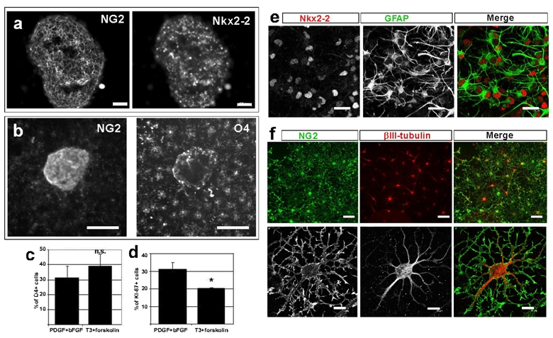
a: Tumor-derived explants in proliferating conditions in presence of both PDGF and bFGF were double immunostained with NG2 and Nkx2.2 antibodies. Bar=50μm.
b: Microscope observation of a glioma-derived explant grown 6 days in culture medium supplemented with PDGF and bFGF and double immunostained with NG2 (green) and O4 (red) antibodies. Bar=200μm
c: Quantification of O4+ cells of 6 day-explant cultures in proliferating (PDGF + bFGF) and differentiating (T3 + forskolin) conditions.
d: Quantification of Ki-67+ cells of 6 day-explant cultures in proliferation and differentiation media. Results show a small decrease of proliferation in differentiating conditions. * Student’s T-Test, P-Value<0.05.
e–f: Tumor cells migrating away from glioma explants in culture medium supplemented with PDGF and bFGF were double immunostained with Nkx2.2 (red) and GFAP (red) antibodies (e), or double immunostained with NG2 (green) and βIII-tubulin (red) antibodies (f). Bar=20μm in e. Bar=50μm in f upper panels and Bar=10μm in f lower panels.