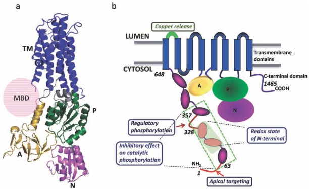
Fig. (1).
Structural organization of human ATP7B. (a) Three dimensional structure of ATP7B showing the transmembrane domains (blue), actuator domain (gold), nucleotide binding domain (pink), phosphorylation domain (green). (b) The regulatory sites which are important for regulation and localization of the protein have been shown. The N-terminal domain (1-648) is the major site for regulation of the molecule. The luminal loop connecting TM1 and 2 (shown in green) is important for copper binding and release (ATP7A).

