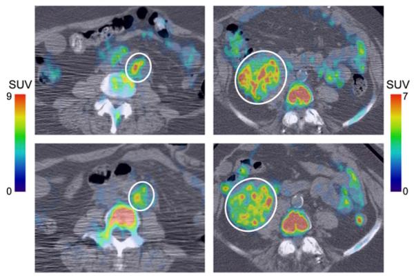
FIGURE 6.

Small vs. large variation in intratumor SUVpeak response. 18F-FLT PET/CT images of periaortic lesion (left, lesion 24) and pelvic tumor (right, lesion 30) at baseline (top) and during treatment (bottom). Lesions are indicated by white circles. Periaortic lesion responded well, exhibiting fairly uniform reduction of 18F-FLT uptake in higher-uptake regions. Consequently, there was little variation in SUVpeak response. In contrast, pelvic tumor responded poorly, with heterogeneous response in higher-uptake regions, resulting in large variation in SUVpeak response.
