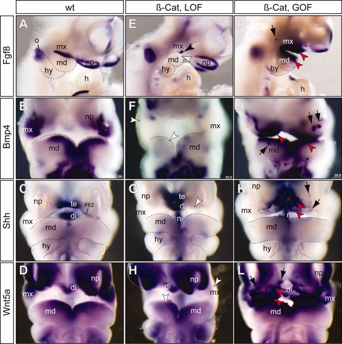
Fig. 5.
β-catenin functions upstream of other epithelial signals. E–L: RNA in situ hybridization (purple color) of embryonic day (E) 10.5 embryos using gene-specific probes indicates decreased expression of Fgf8, Bmp4, Shh, and Wnt5a in the β-catenin loss-of-function (β-cat, LOF) mutants (E–H), while expression of these genes were enhanced in the β-catenin gain-of-function (β-cat, GOF) mutants (I–L). A–D: Wild-type controls. I–L: Patchy ectopic staining (black arrows) of Fgf8 (I), Bmp4 (J), Shh (K), and Wnt5a (L) were observed in the β-catenin GOF mutants. All pictures are frontal views except A, E and I, which are lateral views. di, diencephalon; FEZ, frontonasal ectoderm zone; h, heart; hy, hyoid arch; md, mandibular prominence; mx, maxillary prominence; n, notochord/floor plate; np, nasal placode; o, otic vesicle; te, telencephalon; black arrowhead, residual expression of Fgf8 at the cleft of between mx and md; white arrowhead, reduced expression; red arrowhead, enhanced expression.

