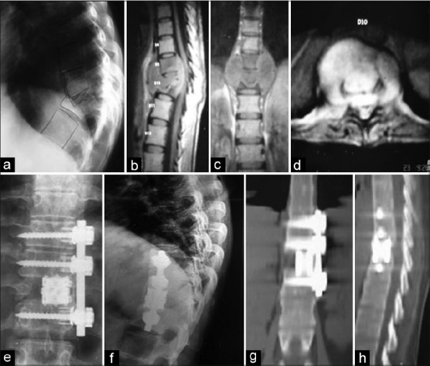
Figure 1.

Preoperative lateral view (a) X-rays of a 26-year-old female with tuberculosis at D9–10 level with kyphosis. Sagittal (b), coronal (c) and axial (d) MRI images of the same patient show vertebral destruction and abscess formation with cord compression. This patient was treated by anterior approach. Postoperative X-ray (e and f) showing good decompression with reconstruction of defect with screw-rod and expandable cage construct. Postoperative CT images (g and h) showing solid bony union at 12 months
