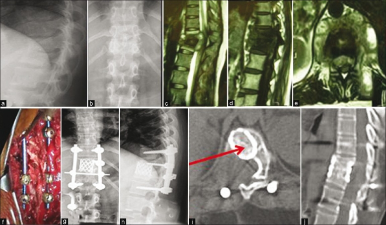
Figure 2.

Preoperative lateral (a) and anteroposterior view X-rays (b) a 32-year-old female with tuberculosis of D11–12. Sagittal T2 WI (c) and T1WI (d) and axial T2WI (e) MRI images show active tuberculosis with abscess formation and cord compression. This patient was treated by posterior extrapleural approach with pedicular screw-rod fixation (f) Postoperative X-rays (g and h) of the same patient show good decompression and kyphosis correction. At 9months followup, solid bony fusion was seen on computed tomography axial and sagittal reconstruction (i and j)
