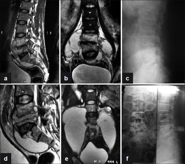
Figure 1.

(a) Sagittal T2WI shows anterior subligamentous spread of large prevertebral collection with interosseous caseation seen in L4, L5 vertebra with endplate erosions and reduced disc height with discitis. (b) Coronal T2WI shows large paravertebral collections with interosseous caseation. (c) Plain X-ray lateral view of lumbosacral spine shows near complete obliteration of L4, L5 disc space with endplate erosions and loss of vertebral height at 3 months. (d, e) T2WI (Sagittal and coronal) show increase in the pre- and paravertebral collections with further loss of vertebral body height of L5 vertebra at 6 months. (f) Plain X-ray AP and lateral views of lumbosacral spine show sharpening of paradiscal cortical margins with better defined disc space and sclerosis of endplates with no paravertebral shadows on final healing
