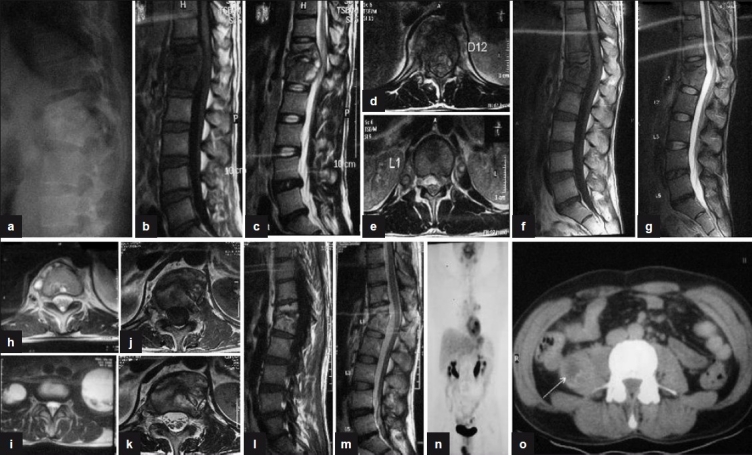
Figure 3.

(a) Plain X-ray lateral view shows anterior wedge collapse of D12 and L1 vetebra with complete obliteration of disc space. (b, c) Sagittal T1W and T2W sections show paradiscal lesions with intraosseous caseation, preserved discs with anterior subligamentous spread of prevertebral collection, and anterior epidural spread of collection compressing the spinal cord. (d, e) T2WI (axial) of D12 and L1 show septate prevertebral collection, intraosseous caseation with large anterior epidural collection, and 60% canal encroachment. (f, g) Sagittal T1WI and T2WI show persistent intraosseous caseation with decrease in epidural collection. (h, i) T2WI (axial) shows persistent septate collections with bilateral psoas collections at 18 months. (j, k) T1WI and T2WI (axial) show near complete resolution of paravertebral collections with residual marrow edema and no epidural extension at 24 months. (l, m) Sagittal T1WI and T2WI show fatty replacement of marrow in D12 and L1 vertebra seen as bright T1 signal with near complete resolution of collections and marrow edema at 30 months. (n, o) Coronal and axial PET scan shows persistent low grade activity in right psoas with no FDG uptake in the vertebra involved
