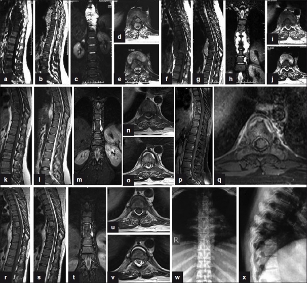
Figure 4.

(a, b) Sagittal T1W and T2W (preoperative) sections show paradiscal lesions with intraosseous caseation, preserved discs with discitis, anterior subligamentous spread of large prevertebral collection, anterior epidural spread of collection compressing the spinal cord. (c) Coronal T2W section shows large paravertebal collection. (d, e) Axial T1W and T2W sections show septate prevertebral collection, intraosseous caseation with large anterior epidural collections, and 80% canal encroachment. (f, g) Sagittal T1W and T2W (4-month post-op) sections show persistent collections and further destruction of D7, D8, and D9 vertebrae. (h) Coronal T2W section shows persistent large paravertebal collection. (i, j) Axial T1W and T2W sections show persistent septate loculated collections and anterior epidural extension. (k, l, m, n, o) MR sagittal, coronal, and axial (at 9 months of second line ATT) sections show significant resolution of collections with persistence of a thin rim of prevertebral collections with preserved discs and near-complete resolution of anterior epidural abscess. (p, q) Post-Gd-DTPA contrast sagittal and axial T1W sections show a thin rim of paravertebral abscess with complete resolution of anterior epidural abscess. (r, s, t, u, v) MR sagittal, coronal, and axial (at 14 months of second line ATT) sections show significant resolution of collections with patchy replacement of bone marrow by fat in D8, D9 vertebrae. A thin rim of anterior prevertebral collection is seen. (w, x) Plain X-ray AP and lateral views (at 18 months of second-line ATT) show sharpening and sclerosis of paradiscal margins with no significant paravertebral shadows suggestive of healing
