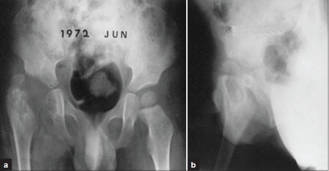
Figure 6A.

X-ray pelvis both hips (a) and (R) hip joint (b) anteroposterior view showing an example of dislocating type of tuberculosis of hip in a 3-year-old boy. Multiple cystic lesions on epiphysis, metaphysis of the proximal femur, and medial aspect of the ischium, with an enlarged acetabulum (mortar type) destructive change are seen, but triradiate cartilage is well preserved
