Figure 6B.

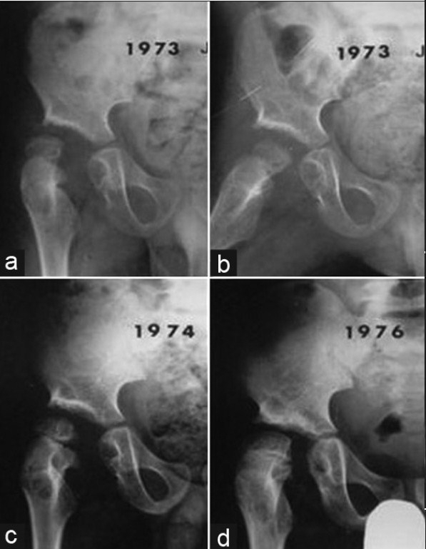
(a, b, c, d) followup radiograms (R) hip of same patient demonstrate the spontaneous gradual medial head migration and disappearance of the cystic bony lesions, though there is slight femoral head subluxation

(a, b, c, d) followup radiograms (R) hip of same patient demonstrate the spontaneous gradual medial head migration and disappearance of the cystic bony lesions, though there is slight femoral head subluxation