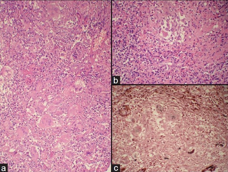
Figure 4.

Photomicrograph showing (a) ill-formed granulomas with many giant cells (H and E, ×200). (b) Inflammatory infiltrate comprising numerous eosinophils and ill-formed granuloma (H and E, ×400). (c) Gomori Methenamine Stain showing septate and acute angle branching fungal hyphae and few spores in the giant cells (GMS, ×400)
