Figure 5.
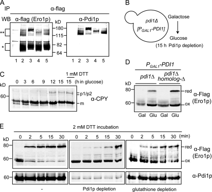
Pdi1p-mediated Ero1 regulation in vivo. (A) Pdi1p forms mixed disulfide with the Ero1p regulatory cysteines. Ero1p-C100A-C105A-Flag and its variants were immunoprecipitated (IP) by anti-flag antibody and analyzed by immunoblots against anti-flag antibody and anti-Pdi1p serum. Lane 1, Ero1p-C100A-C105A-Myc; lane 2, Ero1p-C100A-C105A-flag; lane 3, Ero1p-C100A-C105A-C90A-C349A-flag; lane 4, Ero1p-C100A-C105A-C143A-C166A-flag; lane 5, Ero1p-C100A-C105A-C150A-C295A-flag. The single asterisk indicates a monomer of Ero1p mutants, and the double asterisks indicate mixed disulfides between Ero1p mutants and Pdi1p. WB, Western blot. (B) Pdi1p was depleted by growing pdi1Δ covered with PGAL1-PDI1 in SMM (glucose medium) for 15 h. (C) Immature CPY accumulates as Pdi1p is depleted. A control for fully reduced environment was prepared by treating cells with 1 mM DTT for 1 h. m, mature CPY; p1/p2, nascent and glycosylated CPY, respectively. (D) Oxidation (ox) states of Ero1p after Pdi1p depletion (glucose medium) in a genetic background with pdi1Δ or with pdi1Δ with deletions of all PDI homologs (PDI homolog-Δ). red, reduced. (E) The roles of Pdi1p or glutathione on in vivo activation of Ero1p. Pdi1p-depleted cells (middle) and glutathione-depleted cells (right) were prepared by growing CKY1081 in glucose medium and in galactose medium with 5 mM BSO, respectively. Cells were treated with 2 mM DTT for the indicated time.

