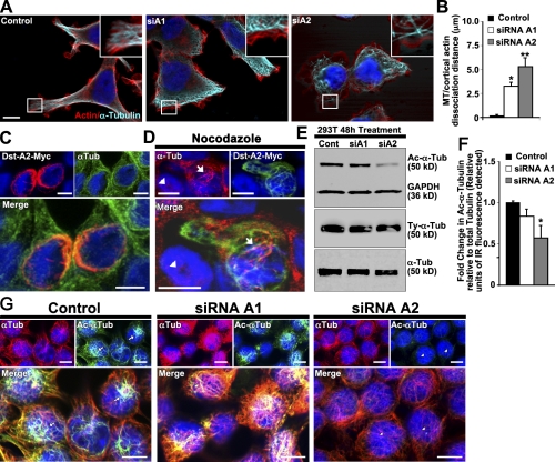
Figure 3.
Loss of dystonin-a2 reduces α-tubulin acetylation status. (A and B) Loss of either dystonin-a1 or dystonin-a2 in 293T cells results in disorganization of the MTs relative to the actin cortex (ANOVA posthoc Dunnett’s t test; *, P < 0.05; **, P < 0.01; n = 9–12). Insets show that MTs do not extend to the actin cortex after dystonin silencing. (C) Dystonin-a2 colocalizes with α-tubulin in the perinuclear region. (D) Overexpression of dystonin-a2–myc stabilizes MTs in 293T cells as indicated by conferring resistance to nocodazole-mediated depolymerization of α-tubulin (compare the protected cell depicted by arrows vs. the susceptible cell depicted by arrowheads). (E–G) 293T Western blot (E) and immunofluorescent analysis (G) of Ac–α-tubulin after dystonin depletion. (F) Quantification of the normalized ratio of Ac–α-tubulin to total α-tubulin showed a significant decrease in Ac–α-tubulin after dystonin-a2 depletion (ANOVA posthoc Dunnett’s t test; *, P < 0.05; n = 4). (G) Note the decrease in Ac–α-tubulin in the perinuclear region of the cell after dystonin-a2 silencing (arrows vs. arrowheads). Error bars show means ± SEM. Tub, tubulin; Cont, control; Dst, dystonin. Bars, 10 µm.

