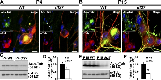
Figure 4.
MT acetylation is reduced in DRGs and primary sensory neurons of dt27J mice. (A and B) P4 (A) and P15 (B) analysis of tubulin acetylation state. Decreased Ac–α-tubulin is evident in dt27J sensory neurons relative to WT in the perinuclear region (arrows vs. arrowheads). (C–F) Western blot analysis of DRG tissue at P4 (C and D) and P15 (E and F) also shows decreased Ac–α-tubulin in dt27J relative to WT samples. Each lane represents DRGs from one animal. (Student’s t test; *, P < 0.05; **, P < 0.01; n = 3). Error bars show means ± SEM. Tub, tubulin. Bars, 10 µm.

