Figure 6.
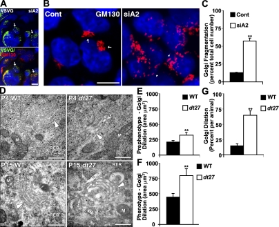
Loss of dystonin-a2 alters Golgi morphology. (A) VSVG localizes with cis-Golgi marker GM130 despite inability to traffic in 293T cells deficient in dystonin-a2. (B and C) Loss of dystonin-a2 in 293T cells promotes Golgi fragmentation (Student’s t test; n = 3). (A and B) Arrows depict compact Golgi, whereas arrowheads depict dispersed Golgi. (D) Electron micrograph of P4 and P15 WT and dt27J DRG sections show dilated Golgi in sensory neurons of dt27J mice. Arrowheads depict the cis-Golgi face, whereas asterisks depict dilated Golgi vesicles. (E–G) Ultrastructural analysis of dt27J Golgi showed increased area at P4 and P15 relative to WT littermates and an increase in the number of dilated Golgi per animal (Student’s t test; n = 4). Error bars show means ± SEM. Cont, control; M, mitochondria; RER, rough ER. **, P < 0.01. Bars: (A and B) 10 µm; (D) 500 nm.

