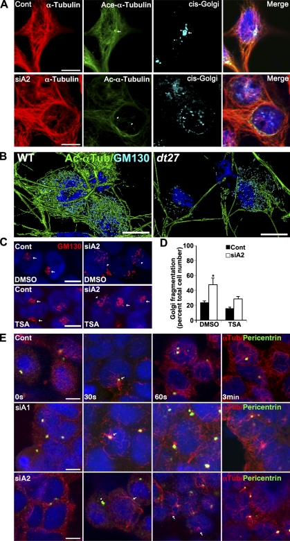
Figure 7.
Impaired MT acetylation results in Golgi fragmentation after loss of dystonin-a2. (A) Silencing of dystonin-a2 in 293T cells results in decreased MT acetylation coupled with reorganization of the Golgi complex. cis-Golgi (GM130) tightly associates with Ac–α-tubulin (arrows vs. arrowheads). (B) 3D rendering of P15 primary sensory neurons from WT and dt27J mice. dt27J sensory neurons show reduced Ac–α-tubulin coupled to a decrease GM130 antigenic labeling in the cell soma. (C and D) Maintenance of MT acetylation status with TSA prevents Golgi fragmentation after depletion of dystonin-a2 (ANOVA posthoc Tukey; *, P < 0.05; n = 6). Arrows depict compact Golgi, whereas arrowheads depict dispersed Golgi. (E) Nocodazole washout of 293T cells after dystonin-a2 depletion shows that MT nucleation from the centrosome is moderately delayed after dystonin-a2 depletion but not dystonin-a1 silencing. Arrows identify sites of MT polymerization. After dystonin-a2 silencing, centrosomal MT polymerization is impeded (arrowhead). Error bars show means ± SEM. Cont, control; Tub, tubulin. Bars, 10 µm.

