Figure 1.
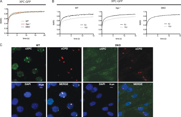
RAD23A and RAD23B immobilize XPC on DNA damage in living cells. (A and B) FRAP analysis of XPC-GFP in the absence and presence of UV damage. The relative fluorescence recovery (It/I0) immediately after bleaching is plotted against the time (seconds). (A) In the absence of DNA damage, no apparent difference in the mobility rate of XPC-GFP could be detected when expressed in either WT, Xpc knockout, or Rad23a/b DKO cells. (B) After UV treatment, part of the XPC-GFP is immobilized (incomplete fluorescence recovery) when expressed in WT cells or in Xpc−/− MEFs. However, when expressed in DKO cells, no immobilization upon UV treatment was observed. Mobilities were measured between 30 and 60 min after UV-C (16 J/m2) treatment. (C) Immunofluorescence analysis of XPC (green channel) at local UV-induced DNA-damaged areas, identified by antibodies that recognize the main UV photoproduct CPD (red channel) in different genetic backgrounds. Nuclei are counterstained by DAPI (blue channel), and the bottom right panel is a merge of all three channels. Cells were fixed 45 min after local UV-C irradiation. (left) Endogenous XPC accumulates at local damaged sites, as is indicated by the presence of the CPDs (red) in WT cells. (right) In Rad23a/b DKO cells, no XPC is found at the local UV damage. Note that the image settings are changed (increased background) to compensate for reduced XPC levels in DKO cells.

