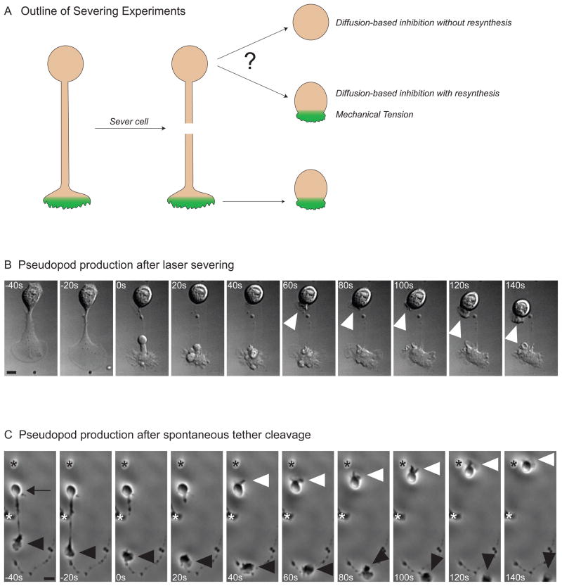
Figure 3. Cells generate a new pseudopod after severing.
A) Outline of severing experiments. Following cell polarization, the pseudopod is removed, and the behavior of the cell body is observed. If the pseudopod had sequestered a non-regenerating limiting component required for polarization, the cell body should not have the material to reanimate. Reanimation of the cell body following severing of the pseudopod would be consistent with short-lived inhibitor generated at the leading edge. This short-lived inhibitor could be due to mechanical tension, a rapidly synthesized limiting component, or a diffusible inhibitor with a short half-life.
B) Pseudopod production after laser severing. DIC images showing a tethered HL-60 that is severed with a laser beam just before 0 seconds. Following severing, the previously quiescent cell body generates a new pseudopod (white arrowhead). The cell body makes a pseudopod after severing in 47% of cells (N = 36). The scale bar is 2.5 microns.
C) Pseudopod production after spontaneous tether cleavage. Phase images of a cell whose pseudopod (black arrowhead) spontaneously breaks free from the cell body (black arrow) at 0 seconds. The cell body makes a new pseudopod within 50 seconds of severing (white arrowhead) and begins to migrate. The asterisks denote neighboring cells. There is significant reanimation of the cell body following spontaneous tether cleavage in 26% of cells (N = 62). The scale bar is 10 microns.

