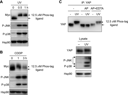
FIGURE 1.
UV irradiation or CDDP-induced mobility shift of YAP. A and B, 293T cells were transfected with Myc-YAP construct, and cells were UV-irradiated (100 J/m2) (A) or treated with CDDP (100 μm) (B) for the indicated times. Cell lysates were analyzed by immunoblotting with the antibodies indicated. C, YAP was immunoprecipitated with anti-YAP antibody from UV-irradiated KB cell lysates. Immunoprecipitates (IP) were incubated in the absence or presence of calf intestinal alkaline phosphatase (AP) together with or without EDTA (50 mm) and immunoblotted with anti-YAP antibody. The arrows indicate the position of YAP in untreated sample, and arrowheads indicate YAP with slow mobility. Hsp90 was blotted as a loading control. For detection of mobility shift of YAP, cell lysates were resolved in polyacrylamide gels containing 12.5 μm Phos-tag ligand when indicated.

