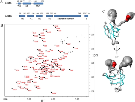
FIGURE 1.
Tertiary structure of the HR domain of OutC. A, schematic representation of the domain organization of inner membrane subcomplex component OutC and secretin OutD. B, assignment of the 1H-15N HSQC spectra for the HR domain. C, sausage representation of the best 20 calculated OutC-HRF3 structures. The secondary structure is shown in color: α-helical residues are in red, and the β-sheet is in cyan. The diameter of the sausage reflects the dynamics of the protein in solution, which is plotted according to the residue α-carbon T2 relaxation profile.

