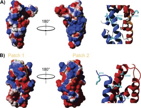
FIGURE 10.
Comparison of the electrostatic surface of Bc28.1 and Bd37. A, two 180° rotated views of the electrostatic surface potential of Bc28.1 calculated using MOLMOL (21), with parameters chosen to represent physiological conditions. Electrostatic potential is represented as a tricolor gradient going from blue (+1 kT) over white (neutral) to red (−1 kT). The dotted yellow ellipse indicates the basic patch on one face of the protein structure. Basic residues accounting for the positive charge of this patch are reported on a zoom in the ribbon representation. B, two 180° rotated views of the electrostatic surface potential of Bd37 calculated using MOLMOL, with parameters as in A. Two basic patches appear clearly at the top of the molecule (dotted yellow ellipses). In the rightmost structure, the basic patch corresponds to the epitope of the monoclonal protective antibody mAb F4.2F8-INT. Basic residues accounting for the positive charge of this later patch are reported on a zoom in the ribbon representation.

