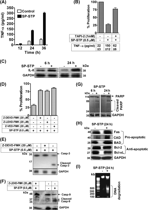
FIGURE 10.
A, quantitation of TNF-α released upon SP-STP addition by ELISA. B, effect of TNF-α inhibitor, TAPI-2, on the SP-STP-induced release of TNF-α from and apoptosis of Detroit 562 cells as revealed by MTT assay. The proliferation of cells treated with the inhibitor alone was taken as control. The table below the graph depicts the amount of TNF-α released (in picograms/ml) from the untreated, SP-STP-treated, and TAPI-2-treated Detroit 562 human pharyngeal cells. C, Western blot analysis showing changes in the expression of IL-8 after 6 and 24 h of treatment. D, effect of caspase inhibitors on the SP-STP-mediated apoptosis of Detroit 562 as determined by MTT assay. E and F, SP-STP-induced cell death is caspase-dependent. Western blots depicting the SP-STP induced cleavage of caspase-3 (E) and caspase-9 (F) in the presence and absence of specific inhibitors. The proliferation of cells treated with the inhibitors alone in each case was taken as control. G, Western blot depicting the cleavage of PARP after 6 and 24 h post-SP-STP addition. H, Western blot analysis showing the changes in the expression of indicated pro- and anti-apoptotic proteins using specific antibodies as described. The GAPDH was used as loading control. I, ethidium bromide-stained 0.8% agarose gel showing DNA fragmentation patterns in SP-STP-treated (+) and untreated (−) pharyngeal cells. The first lane depicts the migration pattern of the DNA molecular weight ladder.

