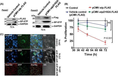
FIGURE 9.
A, Western blot analysis using anti-FLAG and anti-SP-STP antibodies to depict the expression of STP-FLAG (Panel I) and SP-STPD192A (Panel II) 72 h post-transfection in Detroit 562 cell lysates. B, MTT assay depicting the proliferation index of the Detroit 562 cells transfected with pCMV-FLAG (vehicle control), pCMV.stp-FLAG, and pCMV-stpD192A.FLAG at the indicated time points. C, confocal microscopic differential interference contrast (DIC) and merged images showing cytoplasmic localization of GFP (24 h) and nuclear localization of SP-STP-GFP (12 and 24 h). Nucleus is stained with Hoechst33342. Blue DAPI was changed to the red channel for clarity of the superimposed images.

