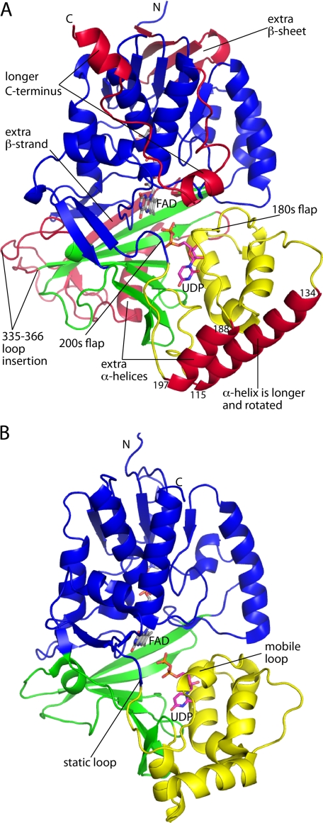
FIGURE 2.
Overall fold of AfUGM. A, structure of the AfUGM protomer. Domains 1, 2, and 3 are colored blue, yellow, and green, respectively. Features that distinguish AfUGM from bacterial UGMs are colored red. B, protomer structure of a bacterial UGM (D. radiodurans UGM, PDB code 3HE3). This figure and others were prepared with PyMOL.

