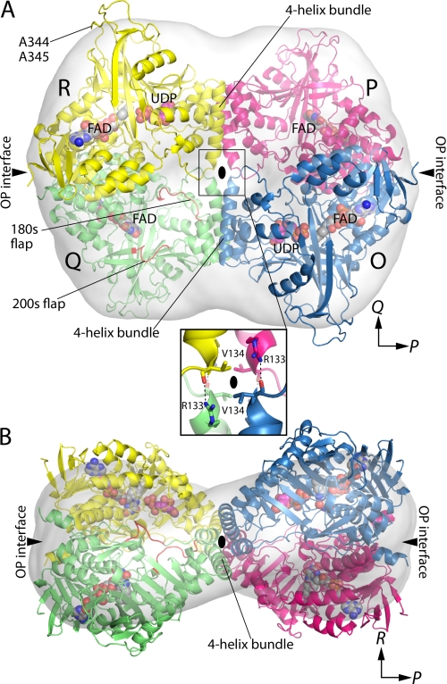
FIGURE 4.
Quaternary structure of AfUGM. A, the tetramer is viewed down the R molecular two-fold axis. Each chain has a different color. The active site flaps of chain Q are colored red. The surface represents the SAXS shape reconstruction. Inset, intersubunit hydrogen bonds at the intersection of molecular two-fold axes. Chains related by the P-axis engage in hydrogen bonding. The oval represents the R two-fold molecular axis. B, the tetramer is viewed down the Q-axis.

