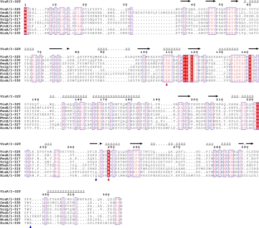
FIGURE 3.
Sequence alignment of known SBPs. The sequences of ViuP from V. cholerae (Protein Data Bank code 3R5S), CeuE from Campylobacter jejuni (PDB code 2CHU), FeuA from Bacillus subtilis (code 2PHZ), YclQ from B. subtilis (code 3GFV), FhuD from E. coli (code 1EFD), FitE from E. coli (code 3BE6), HtsA from Staphylococcus aureus (code 3EIW), and SirA from S. aureus (code 3MWG) were aligned using T-Coffee (28) and prepared using the online program ESPript (29). The secondary structure is represented by α-helix and β-sheet. Blue arrowheads indicate the positions of the basic dyad of ViuP. Red arrowheads indicate the positions of Glu residues of ViuP that may interact with the corresponding ATP-binding cassette transporter ViuDGC.

