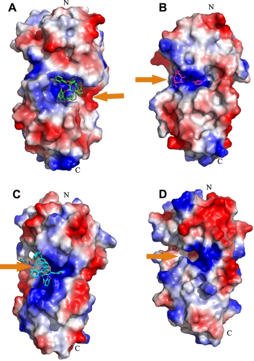
FIGURE 4.
Electrostatic potential map of binding pocket of known catecholate siderophore-binding PBPs. All catecholate siderophore-binding PBPs were displayed with the same orientation of the N- and C-terminal domains. Orange arrows represent the orientation of the binding pocket for ferric vibriobactin. A, ViuP; B, FeuA (Protein Data Bank code 2WHY); C, CeuE (code 2CHU); D, YclQ (code 3GFV) (25–27). The pocket of vibriobactin is on the right side of ViuP, whereas all of the other pockets are on the left side of associated PBPs.

