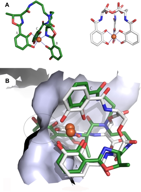
FIGURE 5.
Ferric atom coordination of enterobactin and vibriobactin and superimposed result of [FeIII(Vib)]2− and [FeIII(Ent)]3−. A, blue dashed lines represent the coordination bond. Ferric vibriobactin is on the left, and ferric enterobactin is on the right. [FeIII(Ent)]3− has a 3-fold symmetry axis. B, the white skeleton stick structure is enterobactin, and the green structures are vibriobactin superimposed on enterobactin at the binding site. The encircled region indicates the conflict between [FeIII(Vib)]2− and siderocalin when siderocalin chelates [FeIII(Vib)]2− at the calyx that binds [FeIII(Ent)]3−.

