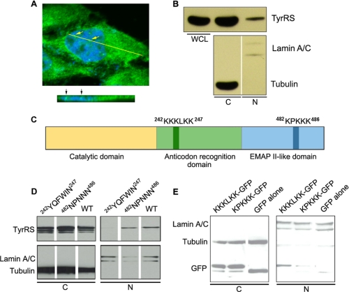
FIGURE 1.
Identification of NLS in human TyrRS. A, confocal immunofluorescence microscopy showing the nuclear distribution of TyrRS in HeLa cells (green, TyrRS; blue, DAPI). The cross section at the yellow line is shown at the bottom. B, HeLa cell fractionation analysis confirming the nuclear distribution of TyrRS. Lamin A/C and tubulin were used as nuclear (N) and cytoplasmic (C) markers, respectively. WCL, whole cell lysates. C, domain structure of human TyrRS and the locations of two potential NLSs. EMAP II, endothelial monocyte-activating protein II. D, mutagenesis and cell fractionation analyses suggesting that KKKLKK is the authentic NLS that directs the nuclear localization of human TyrRS. E, cell fractionation analysis showing that KKKLKK can facilitate the nuclear localization of GFP as a passenger protein.

