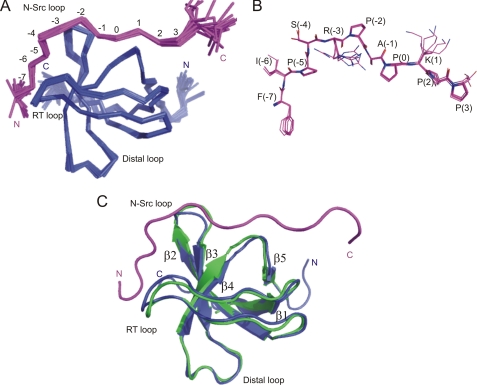
FIGURE 1.
Structure of NbpSH3-Ste20 complex (PDB code 2LCS). A, overlay of the 20 lowest energy structures. The NbpSH3 domain is colored blue and the Ste20 peptide magenta. Well ordered peptide positions are numbered according to Lim et al. (37). B, overlay of five low energy Ste20 peptide conformers in the NbpSH3-Ste20 complex corresponding to peptide positions −7 to 3. C, overlay of the NbpSH3 domain crystal structure (green, PDB code 1YN8 chain B) with the NMR structure of the NbpSH3 domain (blue) in complex with the Ste20 peptide (magenta).

