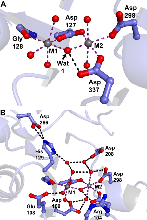
FIGURE 5.
Metal interaction residues in the SUMO-BDP active site. Crystals of SUMO-BDP without added MnCl2 were grown by the hanging drop method in 19% (w/v) PEG 3350, 0.2 m MgCl2, 0.1 m Tris-HCl, pH 8.5, and 20 mm β-mercaptoethanol. A, metal ions. Metal ion 1 (M1) and metal ion 2 (M2), both likely Mg2+ ions, are shown as gray spheres. Side chains from the protein are shown in ball-and-stick format, with oxygen atoms in red, nitrogens in blue, and carbons in light blue. Purple dashed lines show inner-sphere contacts from the protein or water molecules (red spheres) to the metal ions, and black dashed lines show apparent hydrogen bonds. Side chains are labeled with their respective residues, and secondary structure is shown faded for clarity. These coloring and labeling conventions hold for the remainder of the structural figures. B, expanded view of the active site.

