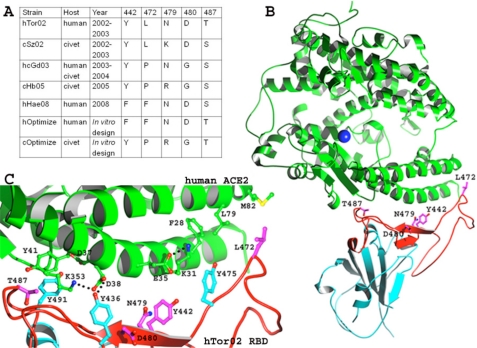
FIGURE 1.
Interface between SARS-CoV RBD and hACE2. A, list of mutations in the RBMs of various SARS-CoV strains. Five representative existent strains and two predicted future strains are defined in the Introduction. B, overall structure of the hTor02 RBD-hACE2 complex (Protein Data Bank code 2AJF). hACE2 is in green, and hTor02 RBD is in cyan (core) and red (RBM). RBM residues that underwent mutations are displayed. C, detailed structure of the hTor02 RBD/hACE2 interface. hACE2 residues are in green, SARS-CoV residues that underwent mutations are in magenta, and SARS-CoV residues that played significant roles in the mutations are in cyan.

