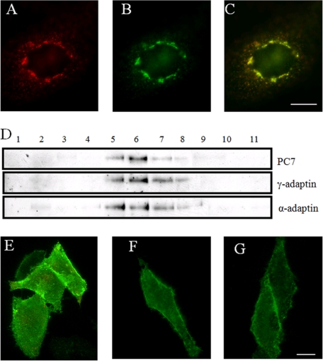
FIGURE 2.
PC7 recycles from PM to TGN via CCVs. A, CHO-K1 cells transfected with f-PC7 were incubated with the anti-FLAG antibody M2 for 30 min at 4 °C and 1 h at 37 °C, fixed, and stained with a TRITC-conjugated secondary antibody to visualize PC7. B, immunofluorescent post-fix staining of the same cells for TGN38 to visualize the TGN. C, merged picture of those cells demonstrates that f-PC7 was transported from the PM to the TGN. D, CCVs were purified using standard procedures as described under ”Experimental Procedures.“ Each fraction from the second density gradient was separated by SDS-PAGE and analyzed by Western blot analysis for α- and γ-adaptin and PC7. E, antibody uptake experiment (30 min 4 °C, 15 min 37 °C) in hypertonic medium on CHO-K1 cells transfected with f-PC7 using the anti-FLAG antibody M2. A double immunofluorescent staining was performed to visualize intracellular PC7 in red and to visualize cell surface-localized PC7 in green. PC7 mainly localizes at the PM under these conditions. F, antibody uptake experiment (30 min 4 °C, 15 min 37 °C) on CHO-K1 cells transfected with f-PC7 and incubated with 30 μm Pitstop 2, using the anti-FLAG antibody M2. A double immunofluorescent staining was performed to visualize intracellular PC7 in red and to visualize cell surface-localized PC7 in green. PC7 mainly localizes at the PM under these conditions. G, CHO-K1 cells transfected with f-PC7 were incubated for 30 min at 4 °C with serum-free medium containing anti-FLAG antibody M2. A double immunofluorescent staining was performed to visualize intracellular PC7 in red and cell surface-localized PC7 in green. PC7 mainly localizes at the PM under these conditions. Scale bar = 10 μm.

