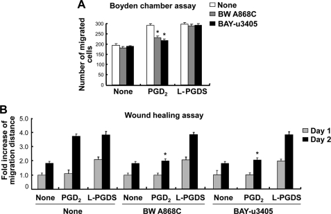
FIGURE 7.
L-PGDS promotes cell migration through a PGD2-independent pathway. NIH3T3 fibroblast cells were treated with the PGD2 (100 ng/ml) or recombinant L-PGDS protein (100 ng/ml) in the presence or absence of BW A868C (DP1-specific receptor antagonist, 10 nm) and BAY-u3405 (DP2-specific receptor antagonist, 10 nm) as indicated. Either the Boyden chamber assay (A) or the wound healing assay (B) was done to evaluate cell migration. The quantification of cell migration was done by either measuring the degree of wound closure (the wound healing assay) or enumerating the migrated cells (the Boyden chamber assay) as described under “Experimental Procedures.” The results are the mean ± S.D. (n = 3). *, p < 0.05; compared with the treatments without antagonists under the same condition.

