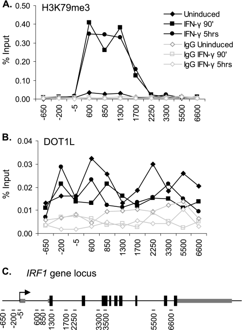
FIGURE 1.
Profile of H3K79me3 and DOT1L at the IRF1 gene. A and B, ChIP using antibodies to H3K79me3 (A) and DOT1L (B) in 2fTGH cells treated with IFN-γ for the indicated times or left untreated. qPCR, using primers spanning the IRF1 gene locus, quantified the precipitate yield reported as the percentage of input. IgG served as the negative control. C, graphic depiction of the IRF1 gene (∼9 kb), showing the location of the qPCR primers (black dashes) used in this study.

