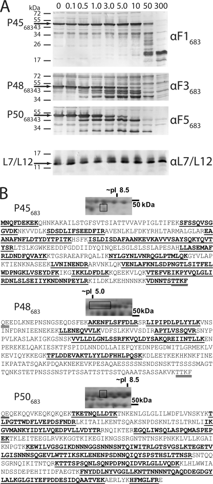
FIGURE 5.
MHJ_0662 (Mhp683 homolog) is present on the surface of M. hyopneumoniae. A, intact and washed (strain J) cells were digested for 15 min with increasing concentrations of trypsin to determine whether P45683, P48683, and P50683 fragments were present on the cell surface. Immunoblots were probed with αF1683, αF3683, and αF5683 sera and identified P45683, P48683, and P50683 fragments of MHJ_0662, respectively. Trypsin concentrations (above blots) are quoted in μg ml−1. Identical lysates were subsequently probed with antiserum to an intracellular control protein (ribosomal protein L7/L12) to confirm cellular integrity. B, surface biotinylation of M. hyopneumoniae. Intact and washed M. hyopneumoniae (strain J) cells were biotinylated and lysed, and biotin conjugates were purified by affinity chromatography. Following separation by two-dimensional electrophoresis, peptides unique to P45683, P48683, and P50683 were identified from purified biotinylated proteins by LC-MS/MS. Underlined sequences indicate peptides identified by LC-MS/MS; M. hyopneumoniae cleavage site motifs are double underlined.

