FIGURE 7.
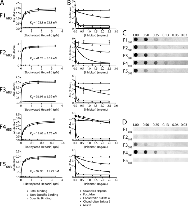
Recombinant proteins F1683–F5683 bind heparin. A, binding of recombinant protein to biotinylated heparin. Total binding (■) was determined by coating recombinant protein F1683–F5683 (10 μg ml−1) to 96-well microtiter plates followed by binding with increasing concentrations of biotinylated heparin (x axis). Nonspecific binding (▴) was determined by binding with unlabeled heparin in a 50-fold excess to biotinylated heparin. Specific binding (◊) was determined by subtracting the nonspecific from the total binding. Kd values for all five recombinant proteins were determined from the specific binding data. B, inhibition of heparin binding by glycosaminoglycans. Recombinant protein (10 μg ml−1) was coated to 96-well microtiter plates and bound with biotinylated heparin in the presence of an increasing excess of inhibitor (x axis). Inhibitors included unlabeled heparin (■), fucoidan (▴), chondroitin sulfate A (▾), chondroitin sulfate B (♦), and porcine type II mucin (●). C, nondenatured recombinant protein dot blots. Recombinant proteins were spotted onto nitrocellulose membrane, blocked, and incubated with 30 μg ml−1 biotinylated heparin. Bound recombinant protein amounts (above blots) are listed in micrograms. D, denatured recombinant protein dot blots. Recombinant proteins were denatured by boiling in an SDS-containing reducing solution before spotting onto nitrocellulose membrane and incubation with heparin as with C. Bound recombinant protein amounts (above blots) are listed in micrograms.

