FIGURE 3.
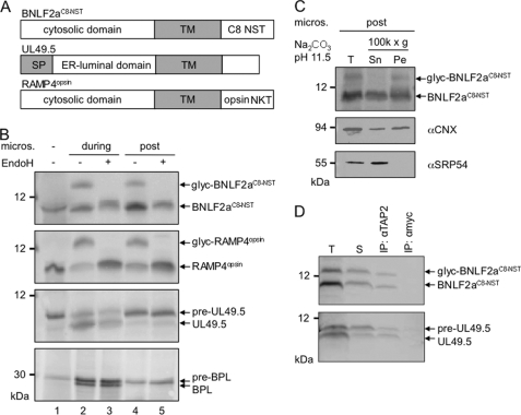
BNLF2a inserts posttranslationally into ER membranes where it binds to TAP. A, schematic illustration of the in vitro-translated proteins BNLF2aC8-NST, UL49.5, and RAMP4opsin. Predicted transmembrane segments (TM) are shown as gray boxes. An N-glycosylation site was added to epitope-tagged BNLF2a and RAMP4. UL49.5 has a signal sequence (SP) that is cleaved by the signal peptidase. B, BNLF2a inserts posttranslationally into the ER membrane. In vitro translation reactions were performed in rabbit reticulocyte lysate in the presence of [35S]Met using templates encoding BNLF2aC8-NST, RAMP4opsin, UL49.5, or bovine prolactin (BPL), respectively. Microsomal membranes were added either at the start of translation (during, lanes 2 and 3) or after termination of translation by puromycin (post, lanes 4, 5). Translation products were analyzed either directly (lane 1, without microsomes) or solubilized (lanes 2-5) and treated with EndoH (lanes 3 and 5). Proteins were separated by Tricine/SDS-PAGE (10%) and visualized by phosphoimaging. glyc, glycosylated protein; pre, preprotein with an uncleaved signal sequence. C, glycosylated BNLF2a is an integral membrane protein. BNLF2aC8-NST was posttranslationally inserted into ER membranes and then subjected to extraction at pH 11.5. Supernatant (Sn), membrane pellet (Pe), and an aliquot before centrifugation (T) were analyzed by SDS-PAGE and visualized by phosphoimaging (BNLF2aC8-NST) or immunoblotting for SRP54 (peripheral membrane protein) and calnexin (CNX; integral membrane protein) as controls. D, posttranslationally inserted BNLF2a interacts with TAP. BNLF2aC8-NST and UL49.5 were in vitro-translated and inserted into TAP-containing Raji microsomes post- or cotranslationally, respectively. After translation, microsomes were collected by sedimentation through a sucrose cushion, solubilized with 2% digitonin, and subjected to immunoprecipitations using antibodies specific for TAP2 or anti-myc (mock). Immune complexes (IP) and 1/20 aliquot of the translation reaction (T) and of the solubilizate (S) were separated by Tricine/SDS-PAGE (10%) and visualized by phosphoimaging. pre-UL49.5, preprotein with signal sequence.

