FIGURE 1.
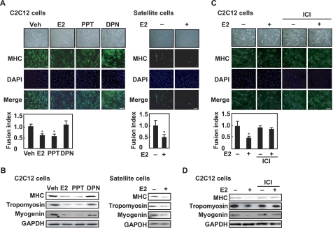
Inhibitory effects of E2 and ERα agonist on myogenesis. A, C2C12 myoblasts (left panel) or satellite cells from young mice (right panel) were cultured in differentiation medium in the presence of E2, PPT, or DPN (each 10 nm) for 8 days. Fixed cells were visualized with anti-MHC antibody and fluorescent-labeled secondary antibody. The nuclei were counterstained with DAPI, and the fusion index was calculated. B, C2C12 myoblasts (left panel) or satellite cells (right panel) were cultured in differentiation medium in the presence of E2, PPT, or DPN (each 10 nm) for 8 days. Cell lysates were subjected to Western blotting with anti-MHC, anti-tropomyosin, anti-myogenin, and anti-GAPDH antibodies. C, C2C12 myoblasts were induced in differentiation medium in the presence or absence of 10 nm E2 and/or 1 μm ICI 182,780 (ICI) for 8 days. Immunoreaction with anti-MHC antibody was performed, followed by incubation with fluorescent-labeled secondary antibody. The nuclei were counterstained with DAPI, and the fusion index was calculated. D, C2C12 myoblasts were induced in differentiation medium in the presence or absence of 10 nm E2 and/or 1 μm ICI 182,780 for 8 days. Cell lysates were analyzed by Western blot with anti-MHC, anti-tropomyosin, anti-myogenin, and anti-GAPDH antibodies. A and C, values are indicated as mean ± S.D. Statistically significant differences compared with the vehicle (Veh) are indicated by *, p < 0.05. Scale bar represents 150 μm. All experiments were performed in triplicate, and each graph is representative of three independent experiments.

