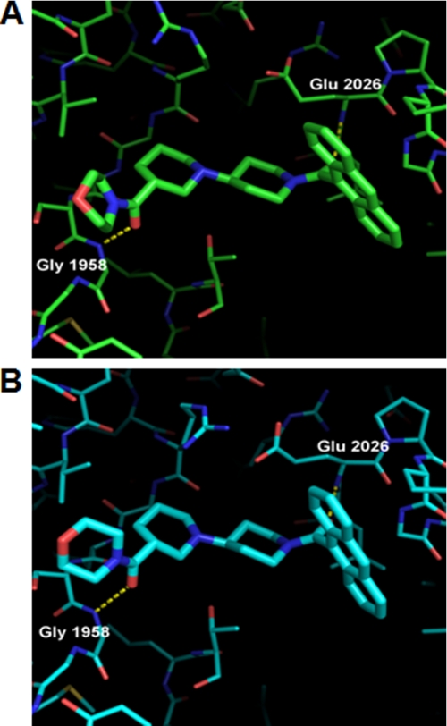
FIGURE 6.
Crystal structure of yCT and yCT-H9 complexed with compound 1. A, binding mode of compound 1 in yCT (green) crystal structure (RCSB entry code 1W2X) shows interactions of the two amide oxygens with the backbone NH of Glu-2026 and Gly-1958 as dashed lines. B, binding mode of compound 1 in the yCT-H9 (cyan) crystal structure, illustrating nearly identical interactions with the active site is shown. Note the shift in position of the terminal morpholine ring (foreground) by nearly 1 Å relative to its position in 1W2X. The structure of compound 1 (PDB code 1W2X) is depicted as green or cyan sticks.

