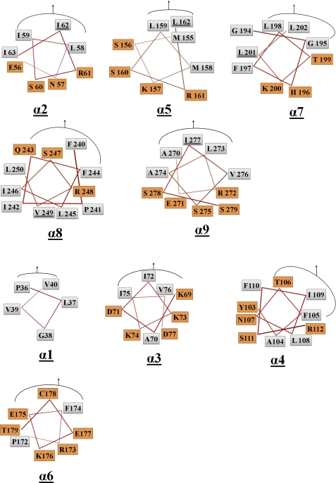
FIGURE 3.
Identification of AHs in TP0453. PyMOL was used to construct helical wheels for all nine AHs based on the closed structures in Fig. 1. For each helical wheel, the hydrophobic and polar amino acids are shown in gray and orange boxes, respectively; the curved line plus arrow denotes the residues facing toward the β sheet. The five AHs (α2, α5, α7–9) indentified by this and our previous analysis are grouped together and correspond to the pink helices in figure 1. Residues selected for mutagenesis are underlined.

