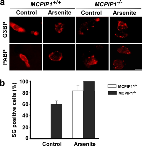
FIGURE 4.
Lack of MCPIP1 results in increase in SG formation. The splenocytes from Mcpip1+/+ and Mcpip1−/− mice were treated with or without arsenite for 1 h. a, SGs were visualized with anti-G3BP and anti-PABP antibodies and anti-mouse secondary antibody labeled with Alexa Fluor 594 (red). Scale bar = 10 μm. b, in G3BP-stained images, the percentage of SG-positive cells was quantified in a two-blind way and is represented as the mean ± S.D. from three independent experiments.

