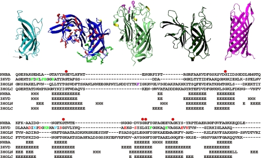
FIGURE 4.
Structural comparison. Top, ribbon representation of proteins with significant structural similarity to NHBA as picked up and aligned by a Dali search (40). From left to right, the C-terminal domain of NHBA, x-ray structure of fHBP (PDB 3kvd), and the N- and C-terminal repeats of TbpB (PDB 3hol) and NspA (PDB 1p4t). The side chains of residues involved in factor H binding (20) or forming a conformational epitope (11) are indicated in red and green, respectively, on the fHBP structure. The side chains of residues involved in transferrin binding are indicated in magenta on TbpB. Bottom, structural sequence alignment of the subfamily containing NHBA, TbpB, and fHBP based on the indicated Protein Data Bank structures. The residues involved in immune response and in factor H and transferrin binding are indicated using the same color coding adopted in the top panels.

