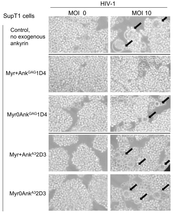
Figure 9.
HIV-1-induced syncytium formation. SupT1/Myr+AnkGAG1D4, SupT1/Myr0AnkGAG1D4, SupT1/Myr+AnkA32D3 and SupT1/Myr0AnkA32D3 were mock-infected (MOI 0; left column) or infected with HIV-1 (MOI 10, right column). Cells were observed at 400X magnification using an inverted microscope. Black arrows point to syncytia.

