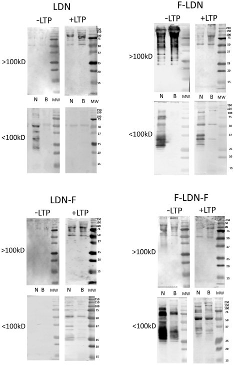
Figure 3. Western/far-Western blot analyses of LDN and (Fucα1,3)-linked LDN glycotopes on plasma proteins.
Fractionated Biomphalaria glabrata plasma (>100 kDa; <100 kDa) from the NMRI (N) and BS-90 (B) snail strains were probed with anti-glycan mABs recognizing LacdiNAc (LDN) and various (Fucα1,3)-linked forms of LDN (F-LDN, F-LDN-F and LDN-F; see Table 1 for glycan structures and mABs used). Blots containing separated plasma proteins were pre-incubated in the presence (+LTP) or absence (−LTP) of S. mansoni larval transformation products (LTP) to determine the effects of LTP exposure on the distribution and intensity of shared glycotopes among proteins of the two B. glabrata strains. Molecular weight markers are indicated on the right.

