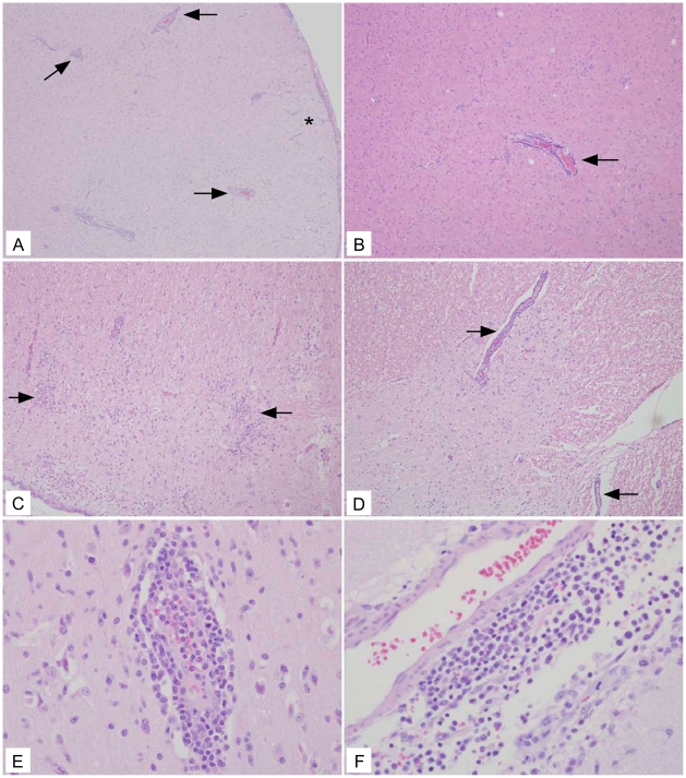
Figure 3. Representative rVSV-wt histology showing lesions in all neural tissue examined.
(A) Frontal cortex (10×) section with severe encephalitic changes including perivascular lymphohistocytic cuffs (arrows) and aggregates of lymphocytes in the neuroparenchyma (*). (B) Frontal cortex (10×) section with perivascular cuff of lymphocytes and histocytes (arrow). (C) Cerebellum (10×) section with aggregates of lymphocytes in the parenchyma (arrows) admixed with increased numbers of reactive glial cells. (D) Spinal cord (10×) section with gliosis admixed with regions of perivascular inflammation (arrows). (E) Frontal cortex (40×) section depicting large numbers of perivascular lymphocytes and histocytes infiltrating into the adjacent gray matter. (F) Basal ganglia (40×) section depicting large numbers of lymphocytes and histocytes both around a meningeal vessel and invading into the adjacent tissue.

