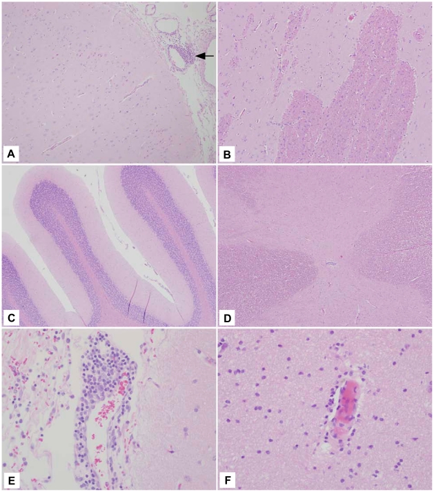
Figure 6. Representative rVSV-MARV-GP histology.
(A) Frontal cortex (10×) section from 59-09 that had a small perivascular cuff of lymphocytes. (B) Frontal cortex (10×) section with no lesions. (C) Cerebellum (10×) section with no lesions. (D) Spinal cord (10×) section with no lesions. (E) Frontal cortex (40×) section with a mild perivascular cuff of lymphocytes. (F) Frontal cortex (40×) section with a scant perivascular cuff of lymphocytes.

