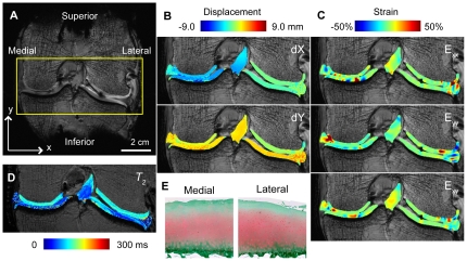
Figure 6. DENSE-FISP for displacements and strains in a complex system inclusive of broad T2 materials.
A human cadaveric tibiofemoral joint was cyclically loaded in the inferior-to-superior direction for DENSE-FISP imaging of a coronal slice (A). Displacements (B) and strains (C) in human tibiofemoral joint show non-homogeneous behavior in the articular cartilage, meniscus, and ligament. DENSE-FISP measures displacements in tissues that range in T2 values (D). Cartilage sections stained with Safranin-O depict some histological signs of degeneration on the medial and lateral femoral condyles (E).

